

Musculoskeletal ultrasound imaging is a non-invasive diagnostic technique that uses high-frequency sound waves to produce images of the muscles, tendons, ligaments, joints, and other soft tissues in the musculoskeletal system. It works by emitting sound waves from a transducer, which are then reflected back and converted into images by the ultrasound machine. The transducer is placed on the skin and moved over the area of interest, allowing the healthcare provider to visualize the internal structures in real-time.
There are several advantages of using musculoskeletal ultrasound imaging compared to other imaging techniques. Firstly, it is a safe and non-ionizing modality, meaning it does not expose the patient to harmful radiation. Additionally, it provides real-time imaging, allowing for dynamic assessment of structures during movement or specific maneuvers. Musculoskeletal ultrasound is also portable and readily available, making it convenient for use in various clinical settings. It is cost-effective and relatively inexpensive compared to other imaging modalities such as MRI or CT scans. Furthermore, it can be used to guide interventions such as injections, providing accurate and precise targeting of the affected area.
In this episode, Erson is joined by Dr. Hannah Cox who recently attended one of his live TMJ Seminars. Upon leaving, she felt prepared to take on the TMJ world! Until that is two days later, she had a patient with high fear avoidance and complaints of open lock TMJ, headaches and neck issues. Luckily, Erson was able to instill her confidence over an online mentoring session and all worked out great over 3 sessions only! Untold Physio Stories is sponsored byHelix Pain Creams - I use Helix Creams in my practice and patients love them! Perfect in combination with joint mobs, IASTM and soft tissue work. Get your sample and start an additional revenue stream for your practice. Click here to get started. https://modmt.com/helixCheck out EDGE Mobility System's Best Sellers - Something for every PT, OT, DC, MT, ATC or Fitness Minded Individual https://edgemobilitysystem.comCurv Health - Start your own Virtual Clinic Side Hustle for FREE! Create your profile in 3 minutes, set your rates, and Curv will handle the rest! From scheduling to payments, messaging, charting, and a full exercise library that allow for patient/clinician tracking, it's never been easier! Click to join Dr. E's new Virtual Clinic Collective to help promote best online practices. Keeping it Eclectic... This article was originally posted on Modern Manual Therapy Blog
.jpg)
Posted by on 2023-05-08
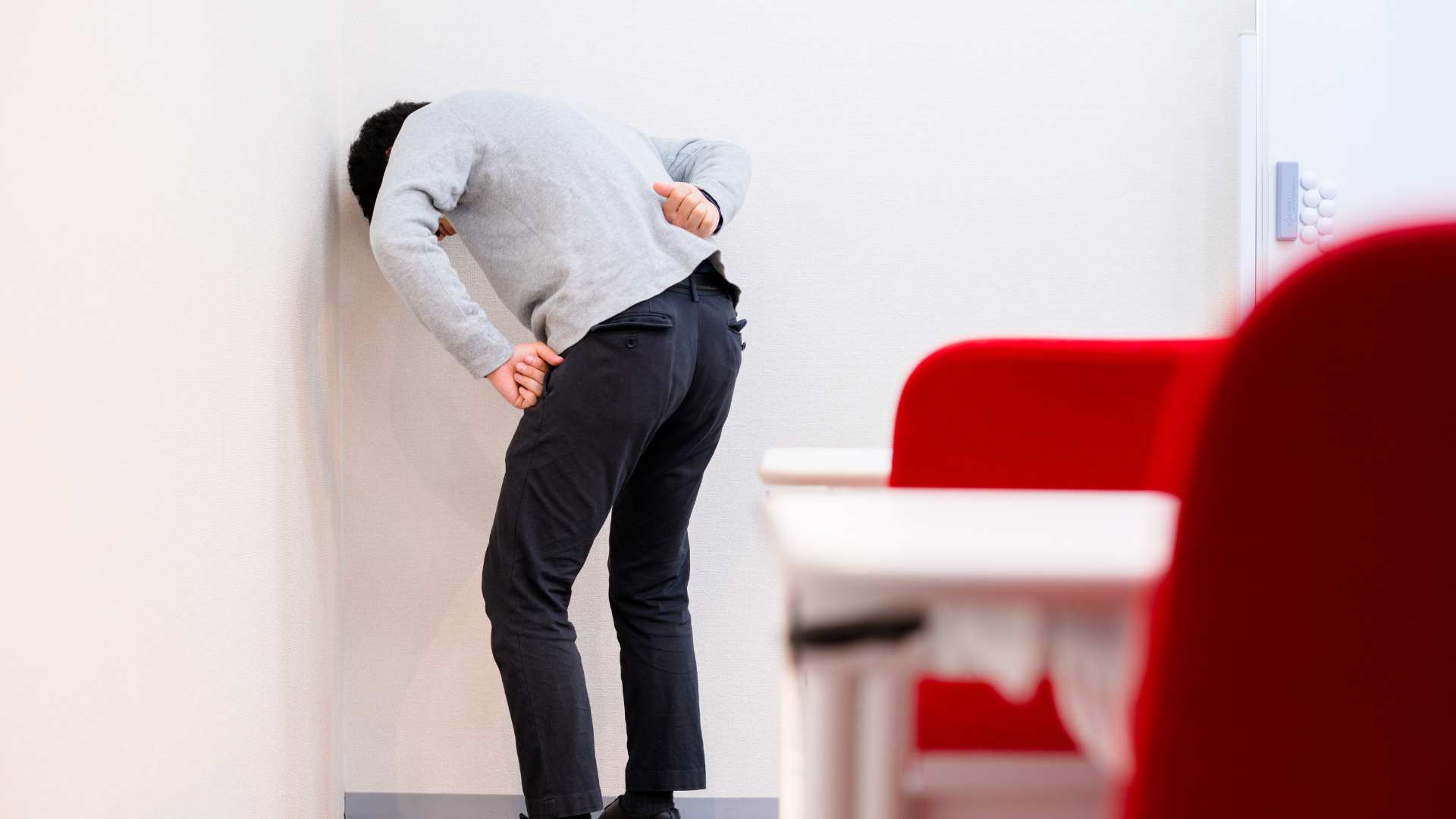
Erson follows up with the difficult lumbar lateral shift patient from this episode a few weeks back. As in the past, he's doing much better and this time Erson takes care not to flare him up! Interestingly enough using the Activforce 2 handheld dynamometer reveals some significant hip and trunk rotation strength percentage differences that could be key to better prevention. Untold Physio Stories is sponsored byHelix Pain Creams - I use Helix Creams in my practice and patients love them! Perfect in combination with joint mobs, IASTM and soft tissue work. Get your sample and start an additional revenue stream for your practice. Click here to get started.Check out EDGE Mobility System's Best Sellers - Something for every PT, OT, DC, MT, ATC or Fitness Minded IndividualCurv Health - Start your own Virtual Clinic Side Hustle for FREE! Create your profile in 3 minutes, set your rates, and Curv will handle the rest! From scheduling to payments, messaging, charting, and a full exercise library that allow for patient/clinician tracking, it's never been easier! Click to join Dr. E's new Virtual Clinic Collective to help promote best online practicesKeeping it Eclectic... This article was originally posted on Modern Manual Therapy Blog
Posted by on 2023-05-04
Introduction SummaryLow back pain (LBP) is a prevalent and costly health problem that affects a significant portion of the global population. Pain developers (PDs) are individuals who are considered a pre-clinical LBP population at risk of developing clinical LBP, which can exact great social and economic costs. Prolonged standing has been identified as a risk factor for LBP, and it is necessary to investigate the risk factors of standing-induced LBP in PDs comprehensively. By identifying these risk factors, appropriate preventive measures can be planned, which may reduce the incidence of standing-induced LBP and its associated costs.This study1 used a systematic review and meta-analysis approach to investigate the distinctive characteristics and risk factors of standing-induced LBP in PDs. The study aimed to identify statistically significant differences between PDs and non-pain developers (NPDs) in demographics, biomechanical, and psychological outcomes and to determine the pooled effect sizes of these differences. The study’s findings have important implications for preventing and managing standing-induced LBP in PDs and for future research investigating the association of these distinctive characteristics to standing-induced LBP and interventions that may modify them.Characteristics of Pain Developers and Non-Pain DevelopersThe systematic review and meta-analysis identified 52 papers and theses involving 1070 participants (528 PDs and 542 NPDs) that were eligible for inclusion. The studies used a prolonged standing duration greater than 42 minutes to classify adult PDs and NPDs without a history of LBP.Significant differences were found between PDs and NPDs in terms of movement patterns, muscular, postural, psychological, structural, and anthropometric variables. PDs exhibited altered motor control in the anterior hip abduction (AHAbd) test and displayed higher lumbar lordosis in individuals over 25 years old. These factors were found to have a statistically significant association with standing-induced LBP.Muscular differences were also identified between PDs and NPDs. PDs had a higher level of co-activation between gluteus medius and the erector spinae muscles, which can lead to increased lumbar loading and potentially contribute to the development of LBP.In terms of postural characteristics, PDs had less trunk control and increased trunk sway during standing compared to NPDs, which may suggest a lack of postural stability.Psychological characteristics were also found to differ between PDs and NPDs. PDs had higher levels of pain catastrophizing, which is the tendency to magnify the threat value of pain and to feel helpless in the face of it, and is associated with increased pain intensity and disability.Finally, anthropometric and structural differences were found between PDs and NPDs. PDs tended to have higher body mass index (BMI) and shorter stature compared to NPDs, which may result in altered spinal loading during standing.These findings suggest that PDs have distinct biomechanical and psychological characteristics that may predispose them to standing-induced LBP. Altered motor control displayed in AHAbd test and higher lumbar lordosis in individuals over 25 years seem to be probable risk factors for standing-induced LBP. The study’s findings have important implications for preventing and managing standing-induced LBP in PDs and for future research investigating the association of these distinctive characteristics to standing-induced LBP and interventions that may modify them.Risk Factors for Standing-Induced Low Back PainThe systematic review and meta-analysis identified several factors that were found to have a statistically significant association with standing-induced LBP:Lumbar fidgets – Participants with PDs displayed more lumbar fidgets, defined as small voluntary or involuntary movements of the lumbar spine, which are indicative of discomfort or pain. This factor was found to have a significant negative effect size (Hedge’s g − 0.72).Lumbar lordosis in participants over 25 years – Participants with PDs had higher lumbar lordosis, defined as the natural curvature of the lumbar spine, in individuals over 25 years old. This factor was found to have a significant positive effect size (Hedge’s g 2.75).AHAbd test – Participants with PDs displayed altered motor control in the AHAbd test, which measures the ability to control the hip and pelvis while lifting one leg. This factor was found to have a significant positive effect size (WMD 0.7).Gluteus medius co-activation – Participants with PDs had higher levels of co-activation between the gluteus medius and erector spinae muscles. This factor was found to have a significant positive effect size (Hedge’s g 4.24).Pain catastrophizing – Participants with PDs had higher levels of pain catastrophizing, which is associated with increased pain intensity and disability. This factor was found to have a significant positive effect size (WMD 2.85).These risk factors suggest that altered motor control, higher lumbar lordosis, increased gluteus medius co-activation, and pain catastrophizing may predispose individuals to standing-induced LBP. The findings may help identify individuals at risk of developing standing-induced LBP and plan appropriate preventive measures.Future research should investigate the association of the reported distinctive characteristics to standing-induced LBP and whether they are manipulable through various interventions. Such interventions may include physical therapy, posture correction, and mindfulness-based stress reduction, among others. Identifying modifiable risk factors may lead to the development of effective interventions for preventing and managing standing-induced LBP in individuals with pre-clinical LBP.Implications for Future ResearchThe systematic review and meta-analysis identified several distinct characteristics and risk factors for standing-induced LBP in PDs compared to NPDs. However, the study authors note that the identified risk factors do not necessarily prove causality or provide a complete understanding of the mechanisms underlying standing-induced LBP. As such, future research should investigate these factors in greater detail, and identify modifiable risk factors that can be targeted for preventive interventions.The study authors recommend that future research should investigate the following areas:Association with standing-induced LBP – Further research should investigate the association of the identified distinctive characteristics and risk factors to standing-induced LBP. Studies should investigate whether these factors are predictive of standing-induced LBP and whether they are specific to standing-induced LBP or generalizable to other types of LBP.Mechanisms underlying standing-induced LBP – Future research should also investigate the underlying mechanisms of standing-induced LBP, such as the interplay between motor control, muscle activation, and posture. Understanding the mechanisms underlying standing-induced LBP can help identify modifiable risk factors and develop effective interventions.Intervention strategies – Future research should investigate the efficacy of various interventions for preventing and managing standing-induced LBP in individuals with pre-clinical LBP. Such interventions may include physical therapy, posture correction, mindfulness-based stress reduction, and other strategies aimed at reducing risk factors identified in this study.Generalizability of findings – Finally, future research should investigate the generalizability of the study findings to other populations, such as individuals with clinical LBP or those with different occupational or lifestyle factors. This will help to determine the applicability of the findings to a broader population and inform the development of preventive measures for standing-induced LBP.ConclusionIn summary, this systematic review and meta-analysis found that pain developers (PDs) – individuals with a history of low back pain (LBP) – have distinct characteristics compared to non-pain developers (NPDs) when exposed to prolonged standing. These characteristics include altered movement patterns, muscular, postural, psychological, structural, and anthropometric variables. The study also identified several risk factors associated with standing-induced LBP, including lumbar fidgets, higher lumbar lordosis in participants over 25 years, AHAbd test, GMed co-activation, and higher scores on the Pain Catastrophizing Scale.These findings have important implications for preventing and managing standing-induced LBP, particularly in individuals with a history of LBP. The study suggests that altered motor control displayed in the AHAbd test and higher lumbar lordosis in individuals over 25 years old are probable risk factors for standing-induced LBP. Therefore, future interventions may focus on improving motor control and reducing excessive lumbar lordosis. Additionally, the study highlights the importance of addressing psychological factors, such as pain catastrophizing, as a potential risk factor for standing-induced LBP.Overall, the study emphasizes the need for a comprehensive approach to preventing and managing standing-induced LBP, including a focus on biomechanical, psychological, and other factors. Future research should investigate the association of these distinctive characteristics to standing-induced LBP and whether they are manipulable through various interventions. By identifying and addressing these risk factors, it may be possible to reduce the prevalence of LBP and improve the quality of life for individuals with a history of LBP.This study emphasizes the importance of developing appropriate preventive measures for standing-induced low back pain (LBP) in pain developers (PDs). PDs are individuals with a history of LBP and are considered a pre-clinical population at risk of developing clinical LBP, which can lead to significant social and economic costs. The study found that PDs have distinct characteristics compared to non-pain developers (NPDs) when exposed to prolonged standing, which suggests that targeted interventions may be necessary to prevent standing-induced LBP in this population.The development of appropriate preventive measures requires a thorough understanding of the risk factors associated with standing-induced LBP in PDs. This study identified several risk factors, including lumbar fidgets, higher lumbar lordosis in participants over 25 years, AHAbd test, GMed co-activation, and higher scores on the Pain Catastrophizing Scale. These risk factors suggest that interventions targeting motor control, lumbar lordosis, and psychological factors may be effective in preventing standing-induced LBP in PDs.In addition to identifying risk factors, the study highlights the importance of comprehensive interventions that address biomechanical, psychological, and other factors associated with standing-induced LBP. These interventions may include postural education, physical therapy, and cognitive-behavioural therapy. By addressing these factors, it may be possible to reduce the prevalence of LBP and improve the quality of life for individuals with a history of LBP.Overall, the study underscores the importance of developing appropriate preventive measures for standing-induced LBP in PDs. Identifying risk factors and developing targeted interventions may help reduce the burden of LBP in this population and improve their overall health and well-being.Dynamic Disc DesignsDynamic Disc Designs offers dynamic anatomical models that musculoskeletal healthcare workers (chiropractors, medical doctors, physiotherapists, osteopaths) can use to help explain how the spine is impacted when one stands, for example. The models are designed to simulate the spinal movement dynamically, allowing various spinal specialists to better illustrate to patients the impact that standing can have on the spine.Using the dynamic disc model, a healthcare worker can demonstrate how the intervertebral discs are compressed when standing due to the force of gravity on the spine. They can show how the discs lose water content and height throughout the day, resulting in reduced shock absorption and increased pressure on the spinal nerves. This can lead to various symptoms, including low back pain, stiffness, and numbness or tingling in the legs. In this particular research highlighted in this post, a practitioner can explain dynamically what excessive lordosis means and how the facets are approximated in this case. Explore.Want to learn in person? Attend a #manualtherapyparty! Check out our course calendar below!Learn more online - new online discussion group included!Want an approach that enhances your existing evaluation and treatment? No commercial model gives you THE answer. You need an approach that blends the modern with the old school. NEW - Online Discussion GroupLive caseswebinarslectureLive Q&Aover 600 videos - hundreds of techniques and more! Check out MMT InsidersKeeping it Eclectic... This article was originally posted on Modern Manual Therapy Blog
![[RESEARCH REVIEW] The High Cost of Standing: Uncovering Risk Factors for Low Back Pain](https://blogger.googleusercontent.com/img/b/R29vZ2xl/AVvXsEgXTsCQGpK-PEaVaLh2d-4MDJt3iZYFUMfzgmUKypDoGEjgXskP71pa-s8bMk_XOK-iWRrL8pLt-vIE6tD_i8NbsgluTbBpfCrbP80CWO3oFOoSZauwQ7U375LUV9hsBh7bwaSz6BJiYSFJfEniuRnDbSGa6swxPr0DzfpYmpWkljZ5TeS2P6031Ioh/s16000/Low%20back%20pain.png)
Posted by on 2023-04-27
Musculoskeletal ultrasound imaging has a wide range of applications in diagnosing musculoskeletal conditions. It is commonly used to evaluate tendon and ligament injuries, such as rotator cuff tears, Achilles tendonitis, and tennis elbow. It can also assess joint inflammation and synovitis, as well as detect and monitor the progression of arthritis. Musculoskeletal ultrasound is useful in diagnosing muscle tears, nerve entrapments, and cysts. It can also aid in the assessment of fractures, bursitis, and soft tissue masses. Overall, it is a valuable tool for identifying and characterizing various musculoskeletal pathologies.
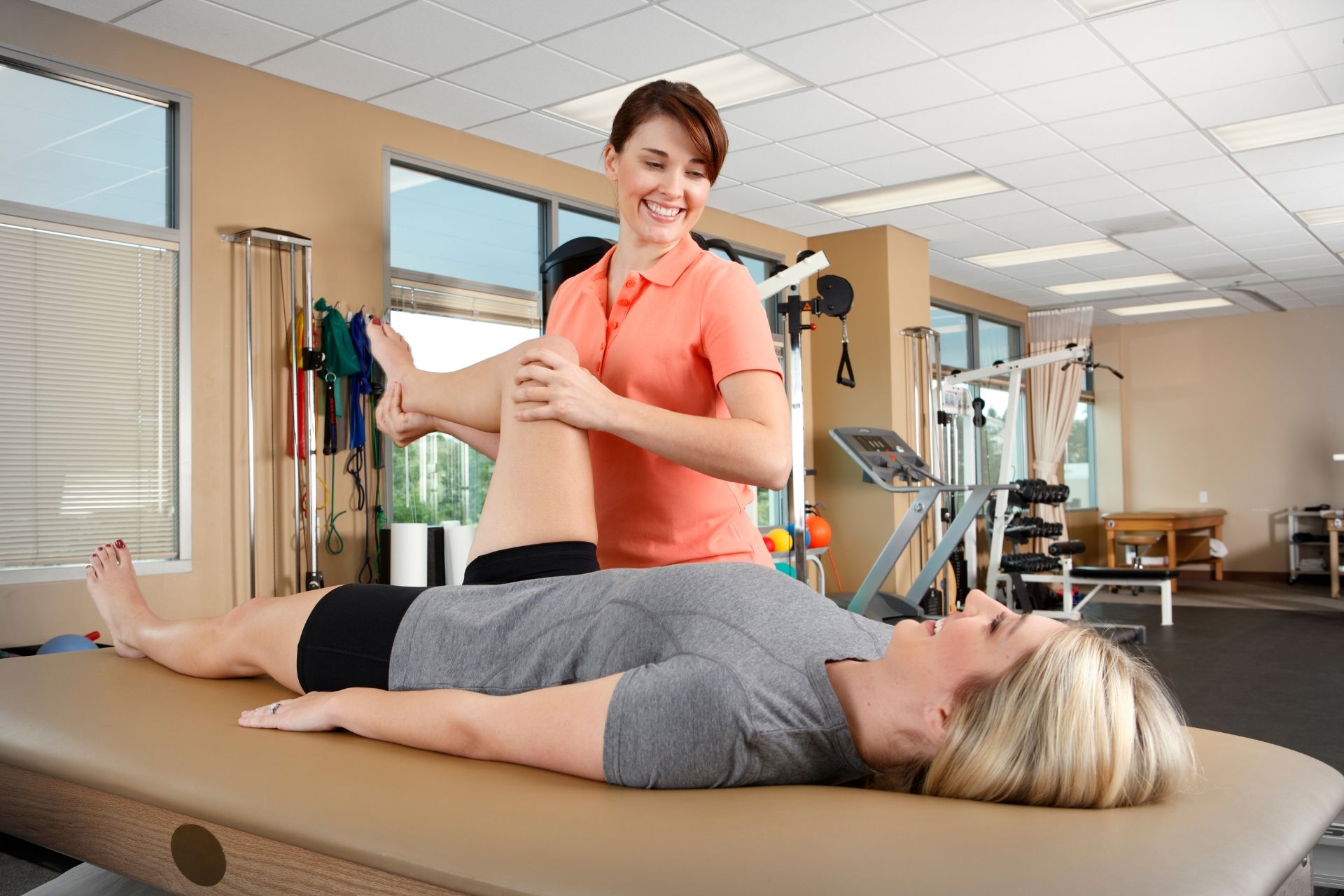
Musculoskeletal ultrasound imaging plays a crucial role in guiding injections and other interventional procedures. By providing real-time visualization of the target area, it allows healthcare providers to accurately guide the needle to the desired location. This ensures that the medication or treatment is delivered precisely to the affected area, maximizing its effectiveness. Musculoskeletal ultrasound can also help identify the presence of fluid collections or cysts, which may need to be aspirated or drained. Additionally, it can assist in performing minimally invasive procedures such as biopsies or aspirations, reducing the need for more invasive surgical interventions.
Despite its many advantages, musculoskeletal ultrasound imaging does have some limitations. One limitation is its operator-dependent nature, as the quality of the images obtained can vary depending on the skill and experience of the sonographer. It may also be limited in obese patients or those with excessive soft tissue, as the sound waves may have difficulty penetrating through the layers. Additionally, musculoskeletal ultrasound has limited penetration compared to other imaging modalities, which may make it less effective in visualizing deeper structures. It may also be challenging to obtain clear images in areas with significant bone or gas interference.

Musculoskeletal ultrasound imaging is generally considered safe and does not have any significant risks or side effects. Unlike other imaging techniques that use ionizing radiation, such as X-rays or CT scans, ultrasound does not expose the patient to harmful radiation. It is a non-invasive procedure that does not require the use of contrast agents, reducing the risk of allergic reactions or kidney damage. However, it is important to note that ultrasound should be performed by trained healthcare professionals to ensure proper technique and interpretation of the images. In rare cases, patients may experience mild discomfort or pressure during the procedure, but this is usually temporary and well-tolerated.
Musculoskeletal ultrasound imaging contributes to the management and treatment of musculoskeletal conditions in several ways. Firstly, it aids in accurate diagnosis and characterization of various pathologies, allowing for appropriate treatment planning. It can help determine the extent and severity of injuries or diseases, guiding decisions regarding conservative management or the need for surgical intervention. Musculoskeletal ultrasound also enables real-time monitoring of treatment progress, allowing healthcare providers to assess the effectiveness of interventions and make necessary adjustments. Additionally, it can assist in guiding rehabilitation exercises and monitoring the healing process. Overall, musculoskeletal ultrasound imaging is a valuable tool in the comprehensive management and treatment of musculoskeletal conditions.
Hydro-massage therapy has been found to be effective in reducing muscle tension and promoting relaxation in pregnant women. The gentle pressure and warmth of the water can help to alleviate muscle tightness and discomfort, providing relief from the physical strain that pregnancy can place on the body. Additionally, the buoyancy of the water can help to reduce the impact on joints and provide a sense of weightlessness, further enhancing relaxation. The rhythmic motion of the water can also have a soothing effect on the nervous system, promoting a sense of calm and reducing stress. Overall, hydro-massage therapy can be a beneficial and safe option for pregnant women seeking relief from muscle tension and a way to relax during this transformative time.
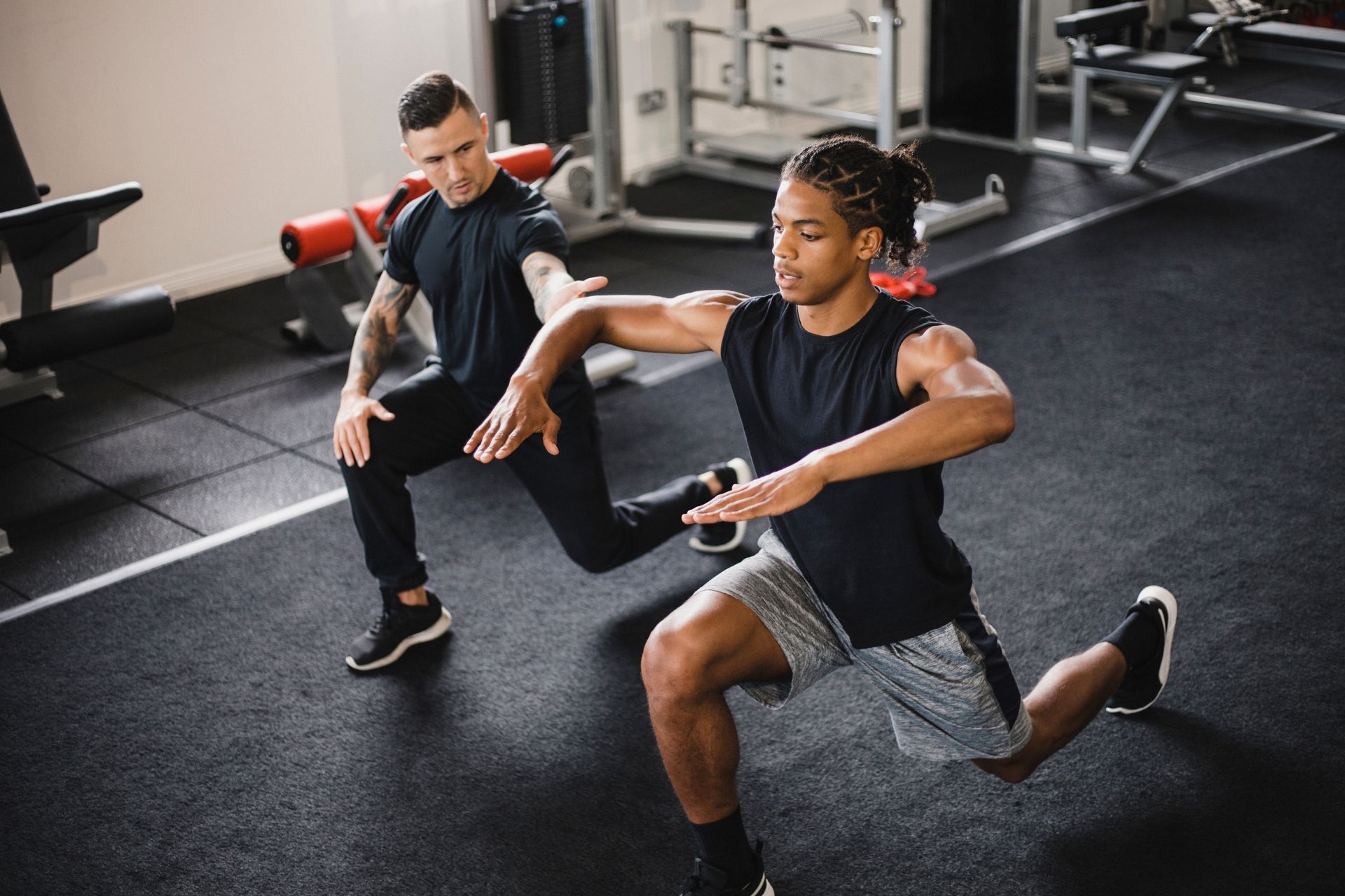
Neuromuscular reeducation plays a crucial role in post-stroke rehabilitation by offering a range of benefits. This therapeutic approach focuses on retraining the brain and muscles to regain lost motor skills and improve overall functional abilities. By incorporating specific exercises and techniques, neuromuscular reeducation helps individuals with stroke-related impairments to enhance their balance, coordination, and proprioception. Additionally, it aids in restoring muscle strength, flexibility, and range of motion, which are often compromised after a stroke. The targeted nature of this rehabilitation method allows for the reestablishment of neural pathways and the promotion of neuroplasticity, facilitating the rewiring of the brain and enabling individuals to regain control over their movements. Moreover, neuromuscular reeducation can contribute to reducing muscle spasticity and preventing secondary complications such as contractures and joint deformities. Overall, this approach empowers post-stroke patients to regain independence and improve their quality of life by optimizing their physical functioning and mobility.
Kinesiology tape can be used as an adjunctive treatment in the management of lymphedema. The indications for using kinesiology tape in lymphedema management include reducing swelling, improving lymphatic flow, enhancing tissue drainage, and providing support to the affected limb. The tape is applied in a specific pattern that creates a lifting effect on the skin, which helps to increase space between the skin and underlying tissues, allowing for improved lymphatic drainage. Additionally, the tape can provide proprioceptive feedback, promoting proper movement and reducing the risk of injury. Overall, kinesiology tape can be a valuable tool in the comprehensive management of lymphedema, helping to alleviate symptoms and improve overall function.