
Common post-operative rehabilitation exercises for knee replacement surgery typically include range of motion exercises, strengthening exercises, and balance and stability exercises. Range of motion exercises may include heel slides, knee bends, and ankle pumps to help improve flexibility and mobility in the knee joint. Strengthening exercises may involve leg lifts, squats, and step-ups to help build strength in the muscles surrounding the knee. Balance and stability exercises may include standing on one leg or using a balance board to improve stability and prevent falls. These exercises are usually performed under the guidance of a physical therapist and can help improve overall function and recovery after knee replacement surgery.
The recovery time for rotator cuff repair surgery can vary depending on the severity of the injury and the individual's overall health. In general, it may take several months to fully recover from this type of surgery. The recommended rehabilitation protocol typically involves a combination of passive and active range of motion exercises, strengthening exercises, and gradual return to functional activities. Passive range of motion exercises may include gentle stretching and assisted movements to help restore flexibility in the shoulder joint. Active range of motion exercises may involve using the muscles of the shoulder to move the joint through its full range of motion. Strengthening exercises may include resistance band exercises, shoulder presses, and rotator cuff exercises to help rebuild strength in the shoulder muscles. The rehabilitation protocol may also include modalities such as heat or ice therapy, electrical stimulation, and manual therapy techniques to help manage pain and promote healing.
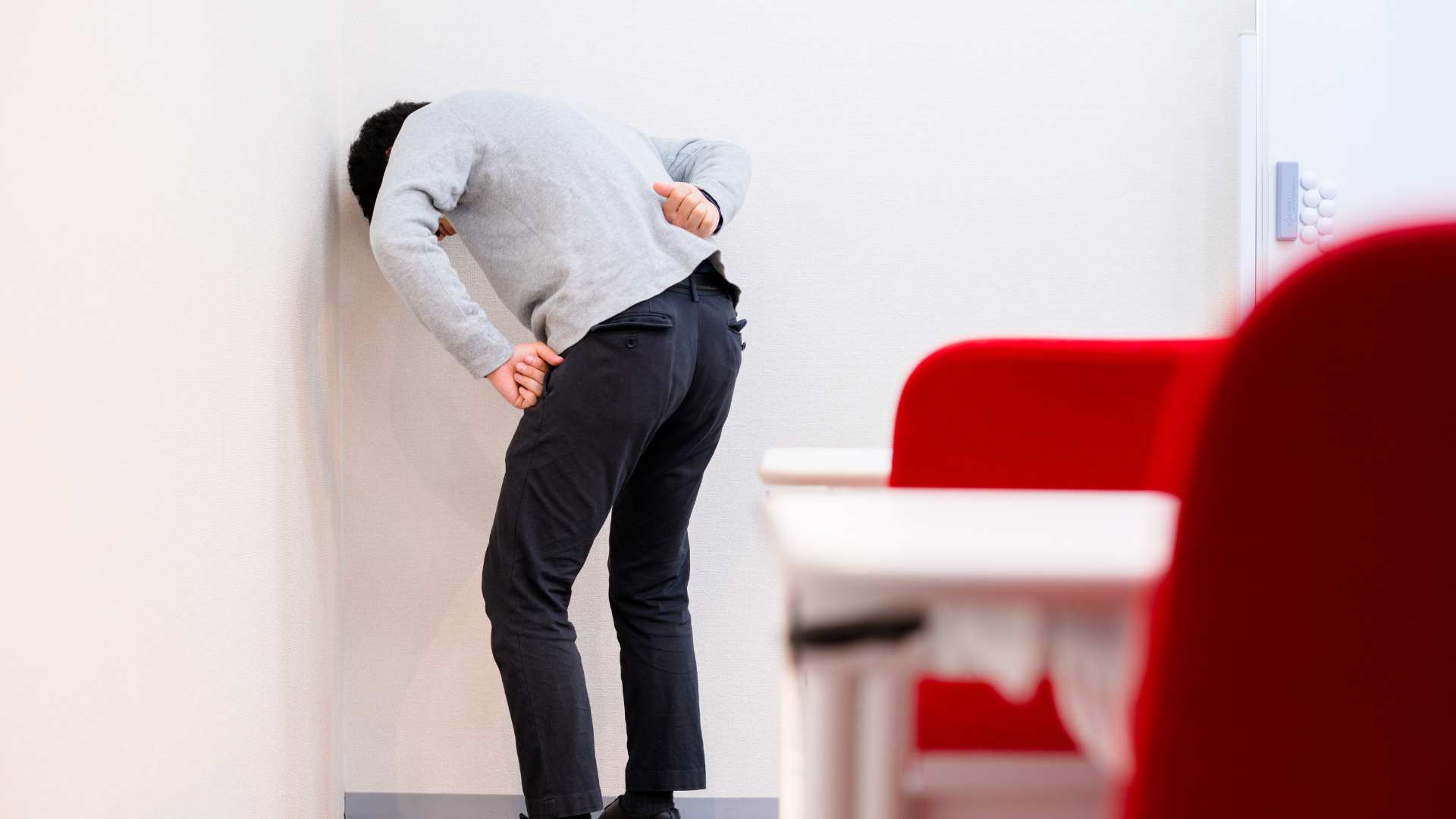
Erson follows up with the difficult lumbar lateral shift patient from this episode a few weeks back. As in the past, he's doing much better and this time Erson takes care not to flare him up! Interestingly enough using the Activforce 2 handheld dynamometer reveals some significant hip and trunk rotation strength percentage differences that could be key to better prevention. Untold Physio Stories is sponsored byHelix Pain Creams - I use Helix Creams in my practice and patients love them! Perfect in combination with joint mobs, IASTM and soft tissue work. Get your sample and start an additional revenue stream for your practice. Click here to get started.Check out EDGE Mobility System's Best Sellers - Something for every PT, OT, DC, MT, ATC or Fitness Minded IndividualCurv Health - Start your own Virtual Clinic Side Hustle for FREE! Create your profile in 3 minutes, set your rates, and Curv will handle the rest! From scheduling to payments, messaging, charting, and a full exercise library that allow for patient/clinician tracking, it's never been easier! Click to join Dr. E's new Virtual Clinic Collective to help promote best online practicesKeeping it Eclectic... This article was originally posted on Modern Manual Therapy Blog
Posted by on 2023-05-04
Introduction SummaryLow back pain (LBP) is a prevalent and costly health problem that affects a significant portion of the global population. Pain developers (PDs) are individuals who are considered a pre-clinical LBP population at risk of developing clinical LBP, which can exact great social and economic costs. Prolonged standing has been identified as a risk factor for LBP, and it is necessary to investigate the risk factors of standing-induced LBP in PDs comprehensively. By identifying these risk factors, appropriate preventive measures can be planned, which may reduce the incidence of standing-induced LBP and its associated costs.This study1 used a systematic review and meta-analysis approach to investigate the distinctive characteristics and risk factors of standing-induced LBP in PDs. The study aimed to identify statistically significant differences between PDs and non-pain developers (NPDs) in demographics, biomechanical, and psychological outcomes and to determine the pooled effect sizes of these differences. The study’s findings have important implications for preventing and managing standing-induced LBP in PDs and for future research investigating the association of these distinctive characteristics to standing-induced LBP and interventions that may modify them.Characteristics of Pain Developers and Non-Pain DevelopersThe systematic review and meta-analysis identified 52 papers and theses involving 1070 participants (528 PDs and 542 NPDs) that were eligible for inclusion. The studies used a prolonged standing duration greater than 42 minutes to classify adult PDs and NPDs without a history of LBP.Significant differences were found between PDs and NPDs in terms of movement patterns, muscular, postural, psychological, structural, and anthropometric variables. PDs exhibited altered motor control in the anterior hip abduction (AHAbd) test and displayed higher lumbar lordosis in individuals over 25 years old. These factors were found to have a statistically significant association with standing-induced LBP.Muscular differences were also identified between PDs and NPDs. PDs had a higher level of co-activation between gluteus medius and the erector spinae muscles, which can lead to increased lumbar loading and potentially contribute to the development of LBP.In terms of postural characteristics, PDs had less trunk control and increased trunk sway during standing compared to NPDs, which may suggest a lack of postural stability.Psychological characteristics were also found to differ between PDs and NPDs. PDs had higher levels of pain catastrophizing, which is the tendency to magnify the threat value of pain and to feel helpless in the face of it, and is associated with increased pain intensity and disability.Finally, anthropometric and structural differences were found between PDs and NPDs. PDs tended to have higher body mass index (BMI) and shorter stature compared to NPDs, which may result in altered spinal loading during standing.These findings suggest that PDs have distinct biomechanical and psychological characteristics that may predispose them to standing-induced LBP. Altered motor control displayed in AHAbd test and higher lumbar lordosis in individuals over 25 years seem to be probable risk factors for standing-induced LBP. The study’s findings have important implications for preventing and managing standing-induced LBP in PDs and for future research investigating the association of these distinctive characteristics to standing-induced LBP and interventions that may modify them.Risk Factors for Standing-Induced Low Back PainThe systematic review and meta-analysis identified several factors that were found to have a statistically significant association with standing-induced LBP:Lumbar fidgets – Participants with PDs displayed more lumbar fidgets, defined as small voluntary or involuntary movements of the lumbar spine, which are indicative of discomfort or pain. This factor was found to have a significant negative effect size (Hedge’s g − 0.72).Lumbar lordosis in participants over 25 years – Participants with PDs had higher lumbar lordosis, defined as the natural curvature of the lumbar spine, in individuals over 25 years old. This factor was found to have a significant positive effect size (Hedge’s g 2.75).AHAbd test – Participants with PDs displayed altered motor control in the AHAbd test, which measures the ability to control the hip and pelvis while lifting one leg. This factor was found to have a significant positive effect size (WMD 0.7).Gluteus medius co-activation – Participants with PDs had higher levels of co-activation between the gluteus medius and erector spinae muscles. This factor was found to have a significant positive effect size (Hedge’s g 4.24).Pain catastrophizing – Participants with PDs had higher levels of pain catastrophizing, which is associated with increased pain intensity and disability. This factor was found to have a significant positive effect size (WMD 2.85).These risk factors suggest that altered motor control, higher lumbar lordosis, increased gluteus medius co-activation, and pain catastrophizing may predispose individuals to standing-induced LBP. The findings may help identify individuals at risk of developing standing-induced LBP and plan appropriate preventive measures.Future research should investigate the association of the reported distinctive characteristics to standing-induced LBP and whether they are manipulable through various interventions. Such interventions may include physical therapy, posture correction, and mindfulness-based stress reduction, among others. Identifying modifiable risk factors may lead to the development of effective interventions for preventing and managing standing-induced LBP in individuals with pre-clinical LBP.Implications for Future ResearchThe systematic review and meta-analysis identified several distinct characteristics and risk factors for standing-induced LBP in PDs compared to NPDs. However, the study authors note that the identified risk factors do not necessarily prove causality or provide a complete understanding of the mechanisms underlying standing-induced LBP. As such, future research should investigate these factors in greater detail, and identify modifiable risk factors that can be targeted for preventive interventions.The study authors recommend that future research should investigate the following areas:Association with standing-induced LBP – Further research should investigate the association of the identified distinctive characteristics and risk factors to standing-induced LBP. Studies should investigate whether these factors are predictive of standing-induced LBP and whether they are specific to standing-induced LBP or generalizable to other types of LBP.Mechanisms underlying standing-induced LBP – Future research should also investigate the underlying mechanisms of standing-induced LBP, such as the interplay between motor control, muscle activation, and posture. Understanding the mechanisms underlying standing-induced LBP can help identify modifiable risk factors and develop effective interventions.Intervention strategies – Future research should investigate the efficacy of various interventions for preventing and managing standing-induced LBP in individuals with pre-clinical LBP. Such interventions may include physical therapy, posture correction, mindfulness-based stress reduction, and other strategies aimed at reducing risk factors identified in this study.Generalizability of findings – Finally, future research should investigate the generalizability of the study findings to other populations, such as individuals with clinical LBP or those with different occupational or lifestyle factors. This will help to determine the applicability of the findings to a broader population and inform the development of preventive measures for standing-induced LBP.ConclusionIn summary, this systematic review and meta-analysis found that pain developers (PDs) – individuals with a history of low back pain (LBP) – have distinct characteristics compared to non-pain developers (NPDs) when exposed to prolonged standing. These characteristics include altered movement patterns, muscular, postural, psychological, structural, and anthropometric variables. The study also identified several risk factors associated with standing-induced LBP, including lumbar fidgets, higher lumbar lordosis in participants over 25 years, AHAbd test, GMed co-activation, and higher scores on the Pain Catastrophizing Scale.These findings have important implications for preventing and managing standing-induced LBP, particularly in individuals with a history of LBP. The study suggests that altered motor control displayed in the AHAbd test and higher lumbar lordosis in individuals over 25 years old are probable risk factors for standing-induced LBP. Therefore, future interventions may focus on improving motor control and reducing excessive lumbar lordosis. Additionally, the study highlights the importance of addressing psychological factors, such as pain catastrophizing, as a potential risk factor for standing-induced LBP.Overall, the study emphasizes the need for a comprehensive approach to preventing and managing standing-induced LBP, including a focus on biomechanical, psychological, and other factors. Future research should investigate the association of these distinctive characteristics to standing-induced LBP and whether they are manipulable through various interventions. By identifying and addressing these risk factors, it may be possible to reduce the prevalence of LBP and improve the quality of life for individuals with a history of LBP.This study emphasizes the importance of developing appropriate preventive measures for standing-induced low back pain (LBP) in pain developers (PDs). PDs are individuals with a history of LBP and are considered a pre-clinical population at risk of developing clinical LBP, which can lead to significant social and economic costs. The study found that PDs have distinct characteristics compared to non-pain developers (NPDs) when exposed to prolonged standing, which suggests that targeted interventions may be necessary to prevent standing-induced LBP in this population.The development of appropriate preventive measures requires a thorough understanding of the risk factors associated with standing-induced LBP in PDs. This study identified several risk factors, including lumbar fidgets, higher lumbar lordosis in participants over 25 years, AHAbd test, GMed co-activation, and higher scores on the Pain Catastrophizing Scale. These risk factors suggest that interventions targeting motor control, lumbar lordosis, and psychological factors may be effective in preventing standing-induced LBP in PDs.In addition to identifying risk factors, the study highlights the importance of comprehensive interventions that address biomechanical, psychological, and other factors associated with standing-induced LBP. These interventions may include postural education, physical therapy, and cognitive-behavioural therapy. By addressing these factors, it may be possible to reduce the prevalence of LBP and improve the quality of life for individuals with a history of LBP.Overall, the study underscores the importance of developing appropriate preventive measures for standing-induced LBP in PDs. Identifying risk factors and developing targeted interventions may help reduce the burden of LBP in this population and improve their overall health and well-being.Dynamic Disc DesignsDynamic Disc Designs offers dynamic anatomical models that musculoskeletal healthcare workers (chiropractors, medical doctors, physiotherapists, osteopaths) can use to help explain how the spine is impacted when one stands, for example. The models are designed to simulate the spinal movement dynamically, allowing various spinal specialists to better illustrate to patients the impact that standing can have on the spine.Using the dynamic disc model, a healthcare worker can demonstrate how the intervertebral discs are compressed when standing due to the force of gravity on the spine. They can show how the discs lose water content and height throughout the day, resulting in reduced shock absorption and increased pressure on the spinal nerves. This can lead to various symptoms, including low back pain, stiffness, and numbness or tingling in the legs. In this particular research highlighted in this post, a practitioner can explain dynamically what excessive lordosis means and how the facets are approximated in this case. Explore.Want to learn in person? Attend a #manualtherapyparty! Check out our course calendar below!Learn more online - new online discussion group included!Want an approach that enhances your existing evaluation and treatment? No commercial model gives you THE answer. You need an approach that blends the modern with the old school. NEW - Online Discussion GroupLive caseswebinarslectureLive Q&Aover 600 videos - hundreds of techniques and more! Check out MMT InsidersKeeping it Eclectic... This article was originally posted on Modern Manual Therapy Blog
![[RESEARCH REVIEW] The High Cost of Standing: Uncovering Risk Factors for Low Back Pain](https://blogger.googleusercontent.com/img/b/R29vZ2xl/AVvXsEgXTsCQGpK-PEaVaLh2d-4MDJt3iZYFUMfzgmUKypDoGEjgXskP71pa-s8bMk_XOK-iWRrL8pLt-vIE6tD_i8NbsgluTbBpfCrbP80CWO3oFOoSZauwQ7U375LUV9hsBh7bwaSz6BJiYSFJfEniuRnDbSGa6swxPr0DzfpYmpWkljZ5TeS2P6031Ioh/s16000/Low%20back%20pain.png)
Posted by on 2023-04-27
Post-operative rehabilitation after spinal fusion surgery carries potential complications and risks. These may include infection, blood clots, nerve damage, and failure of the fusion to heal properly. Infection can occur at the surgical site and may require antibiotics or additional surgery to treat. Blood clots can form in the legs and travel to the lungs, causing a potentially life-threatening condition called pulmonary embolism. Nerve damage can result in weakness, numbness, or pain in the affected area. Failure of the fusion to heal properly, known as nonunion, may require additional surgery to correct. It is important for individuals undergoing post-operative rehabilitation after spinal fusion surgery to closely follow their surgeon's instructions, attend all follow-up appointments, and report any unusual symptoms or complications to their healthcare provider.
Post-operative rehabilitation for individuals who have undergone hip arthroscopy typically focuses on restoring range of motion, strengthening the hip muscles, and improving functional activities. Range of motion exercises may include gentle stretching and joint mobilizations to help improve flexibility in the hip joint. Strengthening exercises may involve exercises such as hip bridges, clamshells, and squats to help build strength in the hip muscles. Functional activities may include walking, stair climbing, and balance exercises to help improve overall function and return to daily activities. Physical therapy may also include modalities such as heat or ice therapy, electrical stimulation, and manual therapy techniques to help manage pain and promote healing.
Recommended exercises and activities for post-operative rehabilitation following ACL reconstruction surgery typically include a combination of range of motion exercises, strengthening exercises, and functional activities. Range of motion exercises may include gentle stretching and joint mobilizations to help restore flexibility in the knee joint. Strengthening exercises may involve exercises such as leg presses, hamstring curls, and squats to help rebuild strength in the muscles surrounding the knee. Functional activities may include walking, stair climbing, and balance exercises to help improve overall function and return to daily activities. It is important for individuals undergoing post-operative rehabilitation after ACL reconstruction surgery to work closely with a physical therapist to ensure proper technique and progression of exercises.
Aquatic therapy can provide several potential benefits in post-operative rehabilitation for individuals who have undergone joint replacement surgery. The buoyancy of water reduces the impact on the joints, allowing for gentle movement and exercise without placing excessive stress on the healing joint. The resistance of the water also provides a low-impact form of resistance training, helping to build strength and improve range of motion. Aquatic therapy can also help reduce pain and swelling, improve circulation, and promote relaxation. Additionally, the water provides a supportive environment, making it easier for individuals to perform exercises and activities that may be challenging on land. Overall, aquatic therapy can be a valuable adjunct to traditional land-based rehabilitation for individuals recovering from joint replacement surgery.
The guidelines for post-operative rehabilitation after total shoulder replacement surgery may vary depending on the surgeon's preferences and the individual's specific needs. However, the general goals of rehabilitation typically include restoring range of motion, strengthening the shoulder muscles, and improving functional activities. Range of motion exercises may include gentle stretching and joint mobilizations to help improve flexibility in the shoulder joint. Strengthening exercises may involve exercises such as shoulder presses, rows, and rotator cuff exercises to help rebuild strength in the shoulder muscles. Functional activities may include reaching, lifting, and carrying tasks to help improve overall function and return to daily activities. Physical therapy may also include modalities such as heat or ice therapy, electrical stimulation, and manual therapy techniques to help manage pain and promote healing. It is important for individuals undergoing post-operative rehabilitation after total shoulder replacement surgery to closely follow their surgeon's guidelines and work closely with a physical therapist to ensure proper technique and progression of exercises.

The Graston Technique offers several advantages for soft tissue mobilization. Firstly, it is a non-invasive and non-surgical approach, making it a safe option for patients. Additionally, it is highly effective in treating various soft tissue conditions such as scar tissue, muscle strains, and tendonitis. The technique utilizes specially designed stainless steel instruments to detect and treat areas of soft tissue dysfunction, allowing for precise targeting and treatment. This targeted approach helps to break down scar tissue, improve blood flow, and promote tissue healing. Moreover, the Graston Technique can be used in conjunction with other therapies, such as exercise and stretching, to enhance overall treatment outcomes. Overall, the Graston Technique provides a unique and effective method for soft tissue mobilization, offering patients a non-invasive and targeted approach to address their specific soft tissue issues.
The principles of tissue healing are essential in guiding treatment interventions. These principles include inflammation, proliferation, and remodeling. In the inflammation phase, the body responds to tissue injury by releasing inflammatory mediators, which attract immune cells to the site of injury. This phase is crucial for removing debris and initiating the healing process. The proliferation phase involves the formation of new blood vessels and the production of collagen, which helps in the formation of new tissue. Finally, the remodeling phase focuses on the reorganization and strengthening of the newly formed tissue. Treatment interventions are guided by these principles, aiming to promote and support each phase of tissue healing. For example, interventions may include the use of anti-inflammatory medications to control excessive inflammation, physical therapy to promote blood flow and tissue regeneration, and exercises to improve tissue strength and flexibility. By understanding and applying these principles, healthcare professionals can optimize treatment interventions and facilitate the healing process.
The McKenzie Method, a widely used approach for the classification and treatment of disc herniation, employs a comprehensive system to assess and manage this condition. The method classifies disc herniation based on its location, size, and direction of protrusion, allowing for a more targeted treatment approach. Treatment typically involves a combination of specific exercises and movements that aim to centralize and alleviate symptoms. These exercises focus on promoting proper spinal alignment, reducing pressure on the affected disc, and improving overall spinal function. Additionally, the McKenzie Method emphasizes patient education and self-management techniques, empowering individuals to take an active role in their recovery and prevent future episodes of disc herniation.
Therapists incorporate mindfulness-based techniques into chronic pain management by utilizing various strategies that promote present-moment awareness and non-judgmental acceptance of pain. They may guide patients through mindfulness meditation exercises, encouraging them to focus their attention on bodily sensations and observe them without reacting or labeling them as good or bad. Therapists may also teach patients to practice mindful breathing, where they pay attention to their breath as a way to anchor themselves in the present moment and cultivate a sense of calm. Additionally, therapists may incorporate body scan exercises, where patients systematically bring their attention to different parts of their body, noticing any sensations or areas of tension. By integrating these mindfulness-based techniques into chronic pain management, therapists aim to help patients develop a new relationship with their pain, reducing suffering and improving overall well-being.
Individuals with chronic obstructive pulmonary disease (COPD) should follow specific exercise guidelines to manage their condition effectively. These guidelines recommend a combination of aerobic exercises, such as walking, cycling, or swimming, along with strength training exercises to improve muscle strength and endurance. It is important for individuals with COPD to engage in regular physical activity to enhance their lung function, reduce breathlessness, and improve overall quality of life. However, it is crucial to consult with a healthcare professional or a pulmonary rehabilitation specialist to develop an exercise program tailored to the individual's specific needs and limitations. The exercise program should be progressive, starting with low-intensity activities and gradually increasing the duration and intensity over time. Additionally, individuals with COPD should incorporate breathing exercises, such as pursed-lip breathing and diaphragmatic breathing, to improve their breathing efficiency and control. Regular exercise, when done correctly and under professional guidance, can significantly benefit individuals with COPD by improving their exercise tolerance, reducing symptoms, and enhancing their overall well-being.